

 Photo by Hans Reniers on Unsplash
The amino acids of insulin were the first proteins ever sequenced in 1955 by English scientist Frederick Sanger. Protein sequencing technologies were the first to be developed. Pehr Edman presented a study in 1950 that revealed a label-cleavage approach for protein sequencing that became known as “Edman degradation.”
During the same period, Frederick Sanger worked on his identification and separation approach, which resulted in insulin sequencing. The 1958 Chemistry Nobel Prize was awarded to Fredrick Sanger for this study. Ten years later, Frederick Sanger was sequencing RNA for the first time, showing the initial form of what is known today as electrophoretic sequencing. To understand the evolution of protein sequencing over the past century, we must first understand what the process entails. So…,
What is the Science Behind Protein Sequencing?
An individual protein or peptide comprises a set of amino acids arranged in a linear order. The amino-terminal (N) end of a protein’s primary structure is usually followed by the carboxyl-terminal (C) end. The design of a peptide can be sequenced or extrapolated from its DNA chronology. The method used to identify and study the amino acid chronology of proteins is known as protein sequencing.
The Edman degradation reaction, mass spectrometry, and protein sequence prediction from the encoding DNA or mRNA sequence, similar to antibody sequencing services, are among the techniques used.
The amino acid arrangement in a protein sequence from amino-terminal to carboxyl-terminal is typically represented as a combination of letters. A code of either one or three letters can act as a tag for each amino acid in the series.
In nature, there exist twenty known amino acids, for example; Glutamic acid (Glu, C), Arginine is a kind of amino acid that is found in (Arg, R), Aspartic acid in (Asp, D), Cysteine (Cys, C), Glutamine is an amino acid found in (Gln, Q), and Asparagine (Asn, N).
What are the Protein Sequencing techniques?
Protein amino acid sequences can be identified using one of 2 well-known techniques. Mass spectrometry is a widely utilized tactic nowadays due to its simplicity. The second approach, Edman degradation with a peptide sequenator, is useful when the protein’s N-terminus has to be characterized.
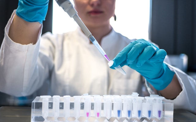 Photo by Julia Koblitz on Unsplash
Mass Spectrometry
A mass spectrometer to sequence proteins has become a widely accepted proteomic approach. The protein is degraded by enzymes and chemicals into a collection of peptides, separated using liquid chromatography with superior efficiency. Each fraction, including up to 10 peptides, is then examined immediately on a multi-analyzer device using a collision-activated dissociation mass spectrometry and liquid secondary ion mass spectrometry.
The research findings involving peptides that are soluble formed via cyanogen bromide and trypsin treatment of apolipoprotein B are made accessible, and the collision-activated dissociation mass spectra are interpreted.
Edman Degradation
The technique of purifying protein by eliminating one residue from the amino end is known as Edman degradation. Pehr Edman devised a novel tagging and cleaving the peptide to overcome the problem of hydrolyzing conditions harming the protein. Edman developed a method for eliminating only one residue while preserving the overall sequence.
This was accomplished by combining Phenyl isothiocyanate with the N-terminal of a phenyl thio carbamoyl derivative. The N-terminal is cleaved under less severe acidic conditions, yielding a cyclic molecule of phenylthiohydantoin PTH-amino acid.
Single-molecule Protein Sequencing
One of the newest theoretical protein sequencing methods involves fixing peptides in a TIRF single-molecule-microscope stage perfusion chamber after covalently tagging with amino acid-specific fluorescent dyes. Each peptide is photographed using TIRF (total internal reflection fluorescence). The N-terminal amino acid is discharged using chemicals by Edman degradation, resulting in a one-amino-acid shorter peptide with a regenerated free N terminus.
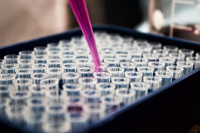 Photo by Louis Reed on Unsplash
The sites of fluorescent dyes within each molecule are revealed by several successions of chemistry (each eliminating one amino acid) and imaging. As illustrated for about three million peptides in the coverslip’s 6.5 mm2 regions, massive amounts of individual peptides can be analyzed simultaneously at suitable attachment densities.
It is feasible to achieve further imaging with a high precision of up to 20 nanometres. Single-molecule localization microscopy makes use of the central location of a single-image molecule established with superior precision than the picture’s size. In the axial (third) dimension, however, achieving the same level of accuracy remains a challenge.
In a TIRF microscope, a photometric approach is possible for decoding the axial location of single molecules. Supercritical Illumination Microscopy Photometric z-Localization with Enhanced Resolution (SIMPLER) doesn’t need hardware changes to a traditional TIRF microscope. It may be used with any two-dimensional single-molecule localization technique to generate three-dimensional images with nanometric accuracy that is almost isotropic.
SIMPLER is immediately usable in just about any experiment using a standard TIRF SMLM microscope. The accessibility of 3-dimensional fluorescence nanoscopy to a broad range of users has ushered in a new age of research into the design and processes of subcellular characteristics and protein-protein interactivity.
In Conclusion
Researchers may use single-molecule protein sequencing to learn more about what, when, where, and why specific targets are expressed, leading to better therapeutic development and outcomes. A comparable possibility exists in research involving neurological and central nervous system illnesses. We believe that single-molecule protein sequencing has a place in the clinical process. We feel that diagnostics have a lot of potential in various fields of medical studies.